


In humans, protozoan parasites cause protozoal infections (or protozoa). These diseases are contagious and threaten with serious complications and consequences. Therefore, timely diagnosis of protozoa requires accurate identification of the pathogen and proper treatment.
What are protozoa parasites
This is a group of single-celled microorganisms that cannot produce nutrients independently. In the process of life, they use other living things to bring them serious diseases. The most common human protozoa parasites are listed below:
- flagella - gardia, leishmaniasis, trichomonas, trypanosoma;
- sarcodal - dysenteric amoeba;
- eyelashes - bursaria, balantidia;
- sporozoans - malaria plasmodium, coccidia, piroplasmas.
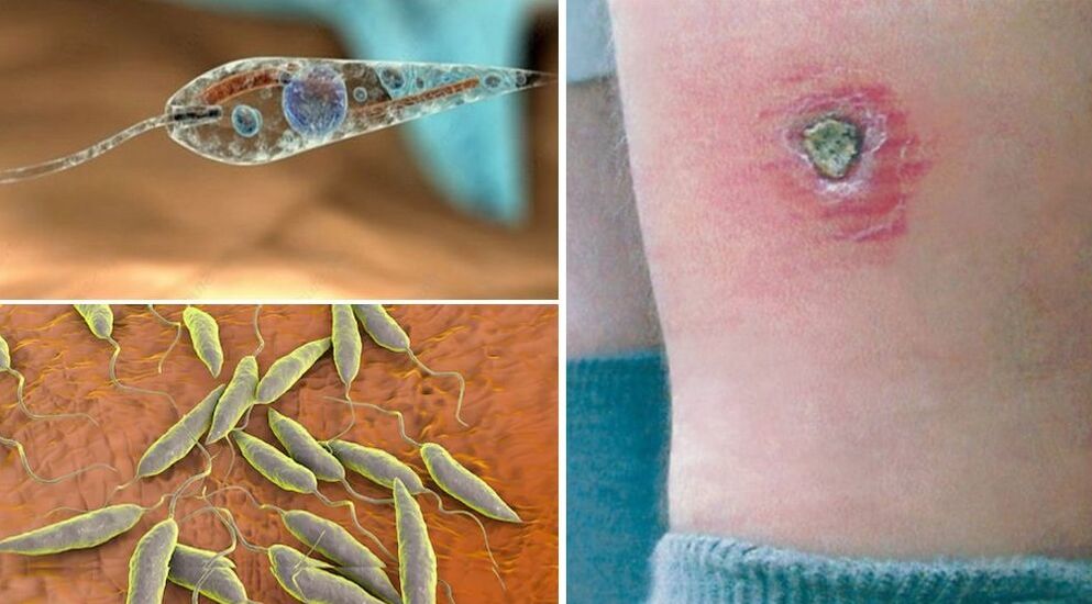
Tripanosome
Life cycle The simplest parasite that occurs in the body of ungulates (antelopes) or infected humans. Carriers are tse-tse flies, which, when bitten by a human, place saliva with protozoa on their skin.
On a note!
Approximately 400 trypanosomes are required for the disease to develop (African trypanosomiasis or sleep disease). With one bite of a tse-tse fly, up to half a million parasites fall.
Features of parasitism and disease:
- tripanosomes first circulate in the blood of an infected person, causing trypanids on the skin (swelling of the face, eyelids), fever up to 40 ° C, swelling of the lymph nodes;
- then single-celled parasites migrate to the cerebrospinal fluid, drowsiness, iridocyclitis, chronic fatigue, lethargy, speech disorders, coordination;
- Advanced forms of trypanosomiasis include limb convulsions, epileptic seizures, nervous and physical fatigue, respiratory paralysis, coma, and death.
Romanovsky-Giemsa test, immunofluorescence test, enzyme immunoassay (ELISA), lymph node puncture are used to diagnose trypanosomiasis. Confirmation of the diagnosis is often made by presenting the blood of a sick person to laboratory pigs. Treatment of sleep disorders involves taking special medications. In the absence of therapy, the patient is likely to be fatal.
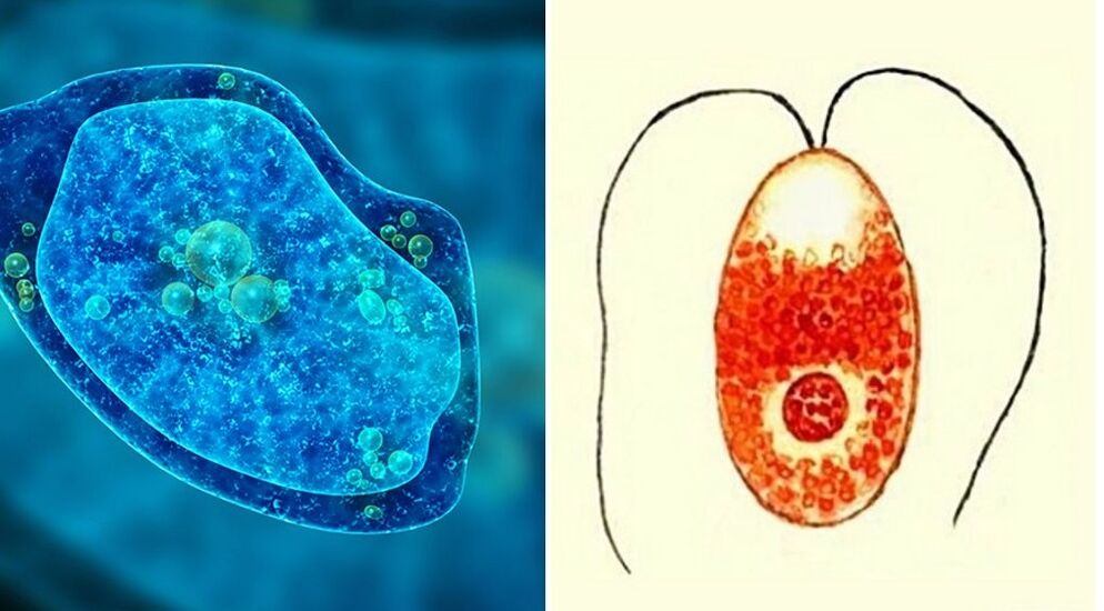
lambliya
Microscopic protozoa (synonyms - giardia or lyamblia) with four pairs of flagella are fully circulating parasites in the human body. Under certain conditions, they cause giardiasis. Giardia attaches to the wall of the small intestine with a large pacifier, often located in the ducts of the liver, gallbladder and other internal organs.
On a note!
Infection with protozoa occurs in food, water, unsanitary conditions. Lymph cysts with embryos remain invasive in the environment for a long time (up to 3 months in fresh water, up to 4 months in sewage). Diagnosis of protozoa is made by microscopy of feces, blood cysts and adults, detection of antibodies on ELISA.
Leishmaniasis
These flagellar protozoa cause leishmaniasis, which is widespread in tropical and subtropical countries. The infection is transmissible - when bitten by the saliva of blood-sucking insects, animals (dogs, ground squirrels). Mosquitoes, gnats, gadflies, ticks can be carriers. There are two types of leishmaniasis in humans:
- skin and mucous skin form (pendinskaya ulcer) - leishmaniasis lives and multiplies on human skin, causing inflammation, swelling, ulcers, trophic ulcers, damage to the respiratory tract;
- Visceral form - leishmaniasis is located in the internal organs (spleen, liver, lungs, heart).
A characteristic feature of cutaneous leishmaniasis is the formation of brown nodules (leishmaniasis) at the site of insect bites. They are then replaced with purulent exudate, round, difficult-to-heal ulcers. The disease lasts 1-2 years, scars remain on the skin. In the visceral form, leishmaniasis causes dysfunction of the adrenal glands, kidneys, liver and spleen. When leishmaniasis is diagnosed, they are found in the bone marrow, lymph nodes, skin fragments, and blood.
On a note!
Treatment of leishmaniasis includes quarantine measures, patient isolation, and drug treatment.
Trichomonas
These are the simplest parasites of the human internal environment, transmitted sexually, through domestic contact or as a result of a congenital infection. Trichomonas has oral, intestinal, and urogenital types. Protozoa are the causative agents of trichomoniasis / trichomoniasis. Urogenital trichomoniasis of the genitourinary system is widespread. The chronic form of the disease is threatened by impotence and persistent infertility. Features of Trichomonas parasitism:
- body size - up to 18 microns, moves quickly thanks to cellular flagella;
- resistant to drugs that determine the chronic course of trichomoniasis;
- dies quickly in the environment, in the air, under direct sunlight;
- stay for a long time on wet towels, sponges, towels, soap dishes;
- frequent infection during vaginal, oral-vaginal intercourse;
- Trichomonas promotes the development of candidiasis, vulvitis, chlamydia, gonorrhea, cystitis.
Diagnosis of trichomoniasis involves the detection of Trichomonas in tampons from the genitals. Treatment involves the use of drugs, treatment with antiseptics. Both partners receive therapy to prevent relapses. Prevention of urogenital trichomoniasis includes recommended measures for all sexually transmitted diseases.
dysenteric amoeba
This sarcoid microorganism is a parasite that causes dangerous diseases in humans. There are two forms of dysentery amoebiasis - intestinal and extraintestinal (liver or lung). The disease begins 7-10 days after infection with symptoms - bloody diarrhea, fever, vomiting.
When left untreated, the consequences of amoebiasis develop - dehydration, exhaustion, weakness, internal bleeding, liver abscess. The infection is most often caused by oral-fecal route. Carriers of amoeba cysts can be insects - flies, gadflies. At the time of diagnosis, tissue forms of protozoa are found in the feces. Treatment of amoebiasis is inpatient, with the use of antibiotics.
Plasmodium malaria
A representative of the simplest spores, the causative agent of a dangerous disease - malaria. The human body serves as an incubator where the parasite's life cycle takes place. Features of parasitism:
- Infection with plasmodium sporozoites occurs during malaria mosquito bites;
- sporozoites enter the bloodstream through the saliva of an infected insect;
- sporozoites are located in the liver, penetrate its cells (hepatocytes);
- where merozoites are formed by mitotic replication;
- when hepatocytes are destroyed, merozoites penetrate erythrocytes;
- gametocytes are formed from merozoites as a result of the sexual cycle;
- when a mosquito is bitten by an infected person, it becomes infected with gametocytes;
- in the mosquito's body, gametocytes migrate to oocysts and then to sporozoites;
- The mosquito infects a healthy person and the cycle repeats itself.
Destruction of erythrocytes and release of gametocytes into the bloodstream in humans is accompanied by fever, vomiting, anemia, convulsions and joint pain. In severe cases, the risk of death increases. Malaria is often relapsing with exacerbations and rest periods. Different protozoa cause three-day and four-day tropical malaria. The main therapeutic and diagnostic agent is quinine - synthesized from cinchona naturally or artificially.
Infusoria balantidia coli
This causative agent of balantidia (or infusor dysentery) lives in the large intestine and causes bleeding wounds in its walls. Infection with protozoa occurs in domestic animals, mainly carriers of pigs. Features of anatomy and parasitism:
- the body of the balantidia is dense, strongly shelled (pellicular) ovary;
- there are many lashes on the surface that serve to move;
- the sexual form of the parasite is necessary for reproduction by simple fragmentation;
- asexual form (cysts) enter the environment with feces;
- The way a person is infected with cysts is through oral feces.
Intestinal placement of protozoa in humans is accompanied by headache, vomiting and dyspepsia. The acute stage of balantidiasis is characterized by fever, signs of severe intoxication, loose stools with blood clots. If not treated in time, a fatal outcome is possible.
Toxoplasma gondii
Microscopic crescent-shaped spores protozoa from the coccidia group are widespread in the environment. They are the causative agent of the disease - toxoplasmosis. Pests inhaled in healthy people are destroyed by immune cells. Features of the disease caused by protozoa parasites in humans:
- often toxoplasmosis is asymptomatic, immunity develops after recovery;
- the parasite affects the visual organs, reproductive, nervous, lymphatic systems, liver, spleen;
- Toxoplasmosis during pregnancy causes severe congenital pathologies in the fetus or its death;
- acute form continues with convulsions, paralysis, hepatic hypertrophy, heart problems;
- In a chronic course, heart dysfunction, damage to the visual organs is possible.
The main owners of protozoa are cats. In their body, giant toxoplasmic colonies are formed from oocysts. People are intermediate hosts and are infected through food, household contact or oral-fecal means.